Anti-Iba1 Microglia Marker
Ionized calcium binding adaptor molecule 1
Ionized calcium binding adaptor molecule 1 or Iba1 is specifically expressed in macrophages and microglia and upregulated during
cell activation. Anti-Iba antibodies for Immunocytochemistry and Western Blot are available and may interest those studying brain injuries and diseases.
Applications
Microglial Marker
Microglial cells are the only brain cells to express Iba-1 (ionised calcium binding adapter molecule 1).
Iba-1 is a 17kDa protein from the large EF hand family of proteins which contain the EF-hand motif.
Iba-1 expression is upregulated in activated microglia enabling differentiation between cells engaged in routine surveillance and those which are activated in response to injury. For this reason, Iba-1, also known as Allograft Inflammatory factor 1 (AIF-1), is often used in immunohistochemistry as a marker for microglia.
Enhanced Iba-1 expression has been observed in traumatic brain injury, ischemia and inflammation.
The Wako Anti-Iba-1 polyclonal antibodies (pAbs) for immunocytochemistry have been raised against a synthetic peptide corresponding to the carboxylterminus of Iba-1, which is conserved amongst human, rat and mouse Iba-1 protein sequences. These antibodies are specific to microglia and macrophages and do not cross react with neurons or astrocytes.
Benefits
Iba-1 (ionised calcium binding adaptor molecule 1) is a specific marker for macrophages and microglia cells.
Suitable for brain development, injury and disease research
Polyclonal rabbit antisera raised against the C terminus of Iba-1
Detection of Iba-1 by immunohistochemistry
Does not cross react with neurons or astrocytes
The original Iba-1 pAb is widely referenced in scientific literature and well suited to double-immunostaining of brain tissue or cell cultures in combination with a monoclonal antibody specific to astrocytes, such as Glial Fibrillary Acid Protein (GFAP).
(Immuno-double staining of rat primary mixed cell culture:
Green: Iba1 Red: Astrocytes reacting with anti-GFAP mAb)

(Data was provided by Department of Neurochemistry, National Institute of Neuroscience (Japan))
| Antigen | Synthetic peptide corresponding to C-terminus of Iba 1 | ||
| Presentation | TBS no preservative | ||
| Conjugate | None | ||
| Species Cross Reactivity | Mouse, Rat, Human | ||

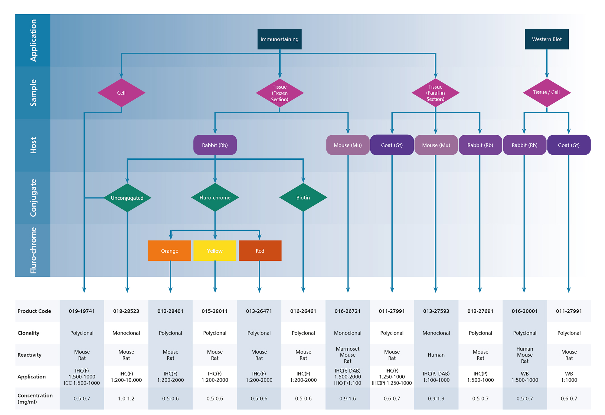
Product Selection